


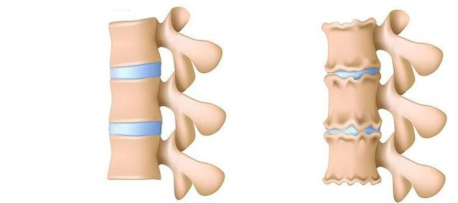
Osteochondrosis of the lumbar region is a disease that deforms and destroys the cartilage of the intervertebral discs in the lower back.Without a layer of cartilage, the distance between the vertebrae is significantly reduced.And with the slightest sharp curves you can shift.The main danger of the disease is the possibility of the formation of Schwandebebralernie.
Can't you lean to lift an object that fell on the floor?Do you suffer acute pain in the lumbar spine and often go, wrap your waist in a warm scarf?Do not ignore the condition that bothers you.
Osteochondrosis of the lumbar region can run for a long time with its duration.There is no need to experience the body for strength.Love your body.And it will expand.
The lumbar region accounts for most of the load from the entire body weight compared to the breast and cervix departments.Therefore, this subspecies of osteochondrosis is most common.
What are the stages of the development of osteochondrosis?
- 1. Level.Excluded.The height of the disc is reduced.A tear is formed in the fibrous ring (the outer layer of the intervertebral disc from the cartilaginous fibers).The lumbar muscles quickly get tired.You feel safe in your back.
- Level 2. Violations of metabolic processes in the jacket of the core (central component of the intervertebral disc, which consists of a cartilage jacket): the cells are dead or completely destroyed.The collagen structure (the protein structure is based on the connective tissue) of the fiber ring is also disturbed.Local pain, a person cannot be dealt with with physical activity, which she previously considered very feasible.
- Level 3. Complete destruction of the fiber ring.Neighboring vertebrae stop being stable.Every unpleasant pose causes pain.Due to the experience of nerve roots that move away from the spinal cord, limbs can become less sensitive and mobile.
- 4. Level.The fabrics of the intervertebral disc become a creepy.The vertebra can turn out in the shell shell.The clinical description depends on individual physiology.
Lumbar pain (lumbago) and pain that the leg sets during the sciatic nerve (Ishias) are one of the most common symptoms that patients seek medical help.Due to the fact that these symptoms are widespread in the general population and their constant growth is also determined, the diagnosis and treatment of such patients on the main activities of the activity of neurosurgical hospitals remains.Despite the widespread pathology, surgical removal of the hernia of the intervertebral disc (MPD) is only required in 10% of patients with the clinical image of the lumbar algia.In the remaining part of the patient, the best effect has conservative treatment, including drug therapy, physiotherapy exercises, the use of physiotherapy treatment methods and a return to the previous daily physical activity.
Disease stages
Degenerative-dystrophic processes begin most often with a deterioration in the shock-absorbing function of the intervertebral disc.
- Determination of blood supply to the intervertebral disc.In adults, the food of the intervertebral discs is carried out by diffusion: blood is only delivered to the vertebrae, and it is already "seeped up" on the windows.In this way, the hard disk is driven for dynamic loads (e.g. walking), since the principle of the pump (drainage of the processed liquid when compressed, the flow of nutrients and oxygen when removing the load).Therefore, the diet of intervertebral discs is particularly difficult under the conditions of a seated lifestyle (hypodynamy).
- Changes in the Pulpic Disk Core.With a deterioration in blood supply, the supply of water, sugar and amino acids is disturbed on the pulpoose nucleus.For this reason, the production of carbohydrates suffers that connect water to water.The core is dehydrated, its structure from gel -like becomes too fibrous, the ability to jump and extinguish shots worsens.This increases the load on the fiber ring and vertebrae, they are rather blocked and injured.
- Changes in the fibrous ring of the intervertebral disc.Due to the flattening of the pulpoose core, the increased load lies on the fibrous disc ring.The fibrous ring loses its strength under conditions of poor blood supply.The instability of the spine occurs, which can lead to the formation of an intermediate remedy, a shift in vertebrae and damage to the spinal cord or nerve roots.
- Storage lead.The formation of Swandromeries.When the fibers of the fibrous ring weaken, the pulp core begins to clear, for example, in the direction of the intervertebral disc channel (disk lead).Such astonishing can continue to break a fiber ring and the formation of a hernia.Read more about the process of the formation of Schwandralernie in a separate article - "Effective treatment of intermediate vertical hernia at home".
- Spondylose is the destruction of the intermediate braler (spondylarrose), the growth of osteofite and the ossification of ribbons.In parallel to the formation of bandrand hernia in osteochondrosis, damage to the band fire joints, destructive changes in the vertebra (cartilage) and league are observed.
As osteochondrosis and the development of complications progress, you have to fall back on medication and increase the doses more and more.This leads to high financial costs and a further deterioration in health due to side effects of medicinal products.
Medicine therapy is usually supplemented by immobilization of a or friend of the spine using orthopedic corset with different degrees of stiffness.
Surgical treatment is only justified in cases in which the compression level of the spinal rook, which is clinically determined, corresponds to the examination, which confirms the breaking of the fiber ring with the "loss" of the hernia of the MPD into the lumen of the vertebral canal [3–6].The results of the surgical treatment in patients with small projections of the hard drive are usually disappointed by doctors and the patient himself.The method for determining a precise diagnosis is magnetic resonance imaging (MRI).About 10% of the people in the community are impossible to carry out routine MRI due to claustrophobia (fear of closed rooms).In this category of people, it is possible to use the "open" MRI in this way with the corresponding loss of the quality of the images obtained.Patients who have previously suffered surgical treatment are necessary to carry out an MRI with contrasting reinforcements in order to delimit the postoperative scar development changes through the true lead of the hard drive.In patients suspected the Hernial lead of the MPD, if the implementation of the MRI is impossible or the results obtained are uninformative, myelography does not acquire a special diagnostic value.
Private diagnostic specialists, who usually interpret the results of the studies, usually exaggerate the degree of damage to the hard drive, since clinical data are compared with "Finds" during tomography.Conclusions such as "Changes correspond to the age of the patient" are almost never found in research protocols.Despite the improvement of neuroimaging techniques, the responsibility for the correctly deceived diagnosis on the clinic's shoulders is, since only he can compare the clinical image with the data obtained during tomography.Increasing the resolution of tomographies easily improved the results of the surgical treatment, but deviations from the norm in asymptomatic patients have been determined.The process of the processes associated with the degenerative determination of the spine in recent years hasmade serious progress.The arthropathy of the vaulted joints is widespread in the general population and is proven to be very common during CT research in people of the middle and older age group.At the same time, pronounced changes in the MPD are not unusual, not accompanied by a break of the fiber ring, but only with a slight "stitch of the pane into the lumen of the vertebral canal or on the side. In some cases, degenerative processes that occur in the MPD can lead to the destruction of the fibrous ring with subsequent breaks, whereby the migration of a part of the pulp core outside of the disk canThe adjoining root of the spinal cord.Koreshka of the nerve of the MPD hernia is caused, beams in pain on the backThe surface of the thigh and the lower leg.An indefinite pain, which is only caused to the buttocks or the thigh area without distribution along the sciatic nerve as well as in bilateral pain in the buttocks or hips that change their location (either right, then left), more often caused by the arthropathy of the arched joints or diffuse degeneration of the MPD.The clinical image of the compression of the Koruska of the MPD hernia can also be a simultaneous pathology (e.g. osteoarthritis of the knee joints).In patients with such pain, surgical treatment will not have the right effect, regardless of which pathology is determined by tomographic examination.In other words, in patients only with the clinic of pain in the back, the removal of the MPD hernia is ineffective, even if tomograms are determined as usual by the lead of the MPD.However, there are also patients in whom the typical image of Ishias is accompanied by a pronounced disabled pain syndrome, while during the studies that were carried out using highly recognizing tomographies, the compression of the roots of the spinal cord is not determined.This category of patients is inappropriate to carry out a surgical intervention, since radicular symptoms generally decrease over time.
It is necessary to introduce yourself to the mechanisms that lead to the development of the pretty mpd in order to recommend patients the volume of permissible movements without forgetting work activity.The forces that contribute to the formation of the hernial advancement are the result of degenerative changes in the MPD and a decrease in the vertical (height) of both the fiber ring and the pulpoose core.The stinging fragment of the MPD in 80% shifts in the posterior direction, while inserting into the lumen of the vertebral canal and in the medial sections of the intermediate hole.This displacement of the Hernie of the MPD in the direction of the center line is facilitated by the holding force of the rear longitudinal band.Up to 10% of the hernial leads are located on the side and spread to the Grandloch (Forsin hernia) or on the outer edge of the hole on which the cerebrospinal spine comes out, causing it to press it, causing it to press it.
In the process of vital activity, dehydration and degenerative changes lead to the loss of the amount of the MPD.These pathological processes contain both a fibrous ring and a pulp core.The more pronounced destruction of the pulpoose nucleus against the background of the simultaneous degeneration of the fiber ring usually only leads to the loss of the MPD without any essential accumulations.With the prevailing changes in the fibrous ring, the vertical forces that affect the pulp core obtained and which affect a derivations of your own weight, as well as the muscles of the back, which act in the side of the side towards the disc, in the lateral direction on the remaining fragment of the pulp, which cannot hold the fiber ring back on the ring.
The summing of these two forces leads to an increase in the centrifugal pressure to the MPD, which together with the stretch component acting on the fiber of the fibrous ring can lead to its break and the fragment of the fragments of the remaining pulp core.After a hernial lead was formed and the "redundant" fragment of the pulp core was outside the fibrous ring, the structure of the MPD is stable again [2].As a result of the forces that influence the degeneratively changed core and fiber ring of the MPD, they are balanced, and their vector, which contributes to a further lead of the fragments of the core, disappears.In some cases, partial degenerative changes in the pulposkern contribute to gas formation within the MPD, followed by excessive pressure on its remaining fragment.The formation of a hernia is also accompanied by the process of gas formation within the disc.
Over the background of the existing degenerative -dystrophic lesion of the spine, excessive and sharp physical activity, which is shown on the patient's back, is usually only a trigger that leads to a detailed clinical image of a compression radicular syndrome, which is frequently and incorrectly viewed by the patient, as the premiere of the lumbar connections.Clinically, MPD hernia can manifest with reflex and compression syndromes.Syndromes are referred to compression in which the upper hernial lead is drawn, pressed and deformed, blood vessels or spinal cord are compressed and deformed.Reflex-reflexes include syndromes, which are caused by the effects of disc hernia on the receptors of these structures, mainly the end of the back-spinal nerves, which leads to the development of reflex and tonic disorders, which are manifested in vasomotor, dystrophic, myofascial disorders.
As mentioned above, surgical treatment with degenerative dystrophic lesion of the Pospinor is only advisable in 10% of the patients, with the remaining 90% react well to conservative measures.The basic principles of using the latter are:
- Relief of pain syndrome;
- Restoration of the correct attitude to maintain the fixation ability of the changed MPD;
- Elimination of muscles and tonic disorders;
- Restoration of blood circulation in roots and spinal cord;
- Normalization of conductivity in nerve fibers;
- Elimination of cicatricial and distance changes;
- Relocation of psycho -Somatic disorders.
Treatment
Today, drugs of the following groups are used in the treatment of osteochondrosis and their complications:
- Net -erous anti -inflammatory medication (NSAIDS) -I form of tablets or injections of medicinal products.These funds can reduce pain and reduce the activity of inflammation.However, the effect of their use does not last long - from several hours to two to three days.Therefore, such funds must be taken for a long time - weeks and sometimes months.At the same time, these drugs have a negative impact on the mucous membranes of the gastrointestinal tract.Your long -term reception is affected by the development of gastritis and ulcerative lesions.In addition, you can negatively influence the work of kidneys, liver and contribute to the development of high blood pressure.At the same time, these funds do not contribute to cleaning slices from dead cells.Therefore, their use is only a way to relieve symptoms for a while, but not to eliminate the main problem.
- CTEPOID (gopmonal) anti -inflammatory medication.As a rule, they are used for severe and impenetrable pain that accompany hernia, radiculitis, Ishias, etc.However, they also negatively influence the mucous membranes of the stomach and intestine, promote the out of the bone and inhibit the production of their own gopmons.And do not contribute to cleaning the focus of dead cells.
- Papasolica are drugs that affect the muscles or nerves that go into the muscles and relax the skeletal muscles.This means that muscle clamps relieve pain for a while, reduce pain and improve blood flow.At the same time, they do not help to clean the tissue of dead cells.Therefore, they do not contribute to healing for osteochondrosis.
- Epiduppal blockade - The introduction of painkillers and gop monal agents in the room between the solid brain cover and the periosteum that covers the vertebrae.It is usually used for intensive pain - in the acute period of intervertebral disc hernia with heavy radiculitis Ishias.Depending on the composition, such an injection helps to relieve pain for several hours to several days.After the expiry date, the manifestations of the disease are returned, since the procedure does not help to restore the metabolic processes in discs.In addition, there is a risk of violation of blood vessels and nerves in the execution.
Conservative treatment methods include various orthopedic effects on the spine (corset real estate, traction, manual therapy), physiotherapy (therapeutic massage, physiotherapy exercises, acupuncture, electrotherapy, mudding, various types of warming), paravertebral, peridural blockade and drug therapy.The treatment of the degenerative dystrophic lesion of the spine should be complex and gradually.As a rule, the general principle of conservative measures is the appointment of analgesics, non -steroidal anti -inflammatory drugs (NSAIDS), muscle relaxants and physiotherapy.
The analgesic effect is achieved by the appointment of Diclofenac, KetoProfen, Lornoxicam, Tramadol.Loroxes, which are available in both injection and tablet forms, has a pronounced analgesic and anti-inflammatory effect.
NSAIDs are the most frequently used medication for degenerative dystrophic damage to the spine.They have an anti -inflammatory, analgesic and antipyretic effect that is associated with the suppression of the enzyme cycloxygenase (COC -1 and TSOS -2) that regulates the conversion of arachidonic acid into prostaglandins, prostacillas, thromboxans.In older people and patients with risk factors for side effects, it is advisable to carry out the "cover" of the gastrotectors under the "cover".In such patients, after completing the injection therapy of NSAIDs, the transition to the tablet forms of the COO -2 inhibitors, which have a lower severity of the side effects of the stomach intestine, is advisable.
In order to eliminate pain associated with an increasing muscle tone, it is advisable to include central musclexants in complex therapy.
The surgical treatment of the degenerative dystrophic lesion of the spine is justified with the ineffectiveness of complex conservative measures (within 2 to 3 weeks) in patients with hernias of MPD (usually more than 10 mm) and non -can -channel radical symptoms.There are emergencies for surgical intervention with a "fallen" sequestra in the lumen of the vertebral canal and expressed compression of the roots of the spinal cord.The development of the caudal syndrome is facilitated by acute radiculomil cemy, which leads to severe hyperalgic syndrome, if even the prescription of drug analgesics does not reduce the use of blockade (with glucocorticoid and anesthetic).It is important to note that the absolute size of the pane hernia does not have a fixed value for the final decision on surgical interventions and should be taken into account in connection with the clinical image and is determined by tomographic examination.In 95% of cases, open access to the vertebral canal is used in the hernia.At the moment, various discopation techniques (cold plasma coagulation, laser reconstruction, etc.) have not been carried out at the moment, and their use is only justified for the requirements of the MPD.The classic open microsurgical removal of the hernia of the disc is used using microsurgical tools, binoculars or an operational microscope.The analysis of the distance treatment results (within more than 2 years) 13,359 patients who had undergone the removal of the MPD hernia, 6135 of whom the sequestral was removed, and 7224 aggressive discsctomy was shown that the relapse of the pain is 2.5 -more often (27.8% against 11.6%).Times more likely (7% compared to 3.5%) in patients who only remove sequestration.The quality of life is more reduced in patients with pain syndrome, while repeated hernia formation is not always clinically manifested.
In summary, I would again emphasize the need for a thorough clinical examination and analysis of tomograms in order to make an optimal decision on the selection of the tactics for the treatment of a specific patient.